Our understanding of the living world is only as good as our instruments for measuring it. Across my career, I’ve worked with many modalities of imaging and spectroscopy - vibrational and electronic, optical and electron, micro and macro. I build these hardware systems to capture photons from life’s interaction and processes, and the software to interpret their results. Here are some of the systems I’ve validated, built, or deployed.
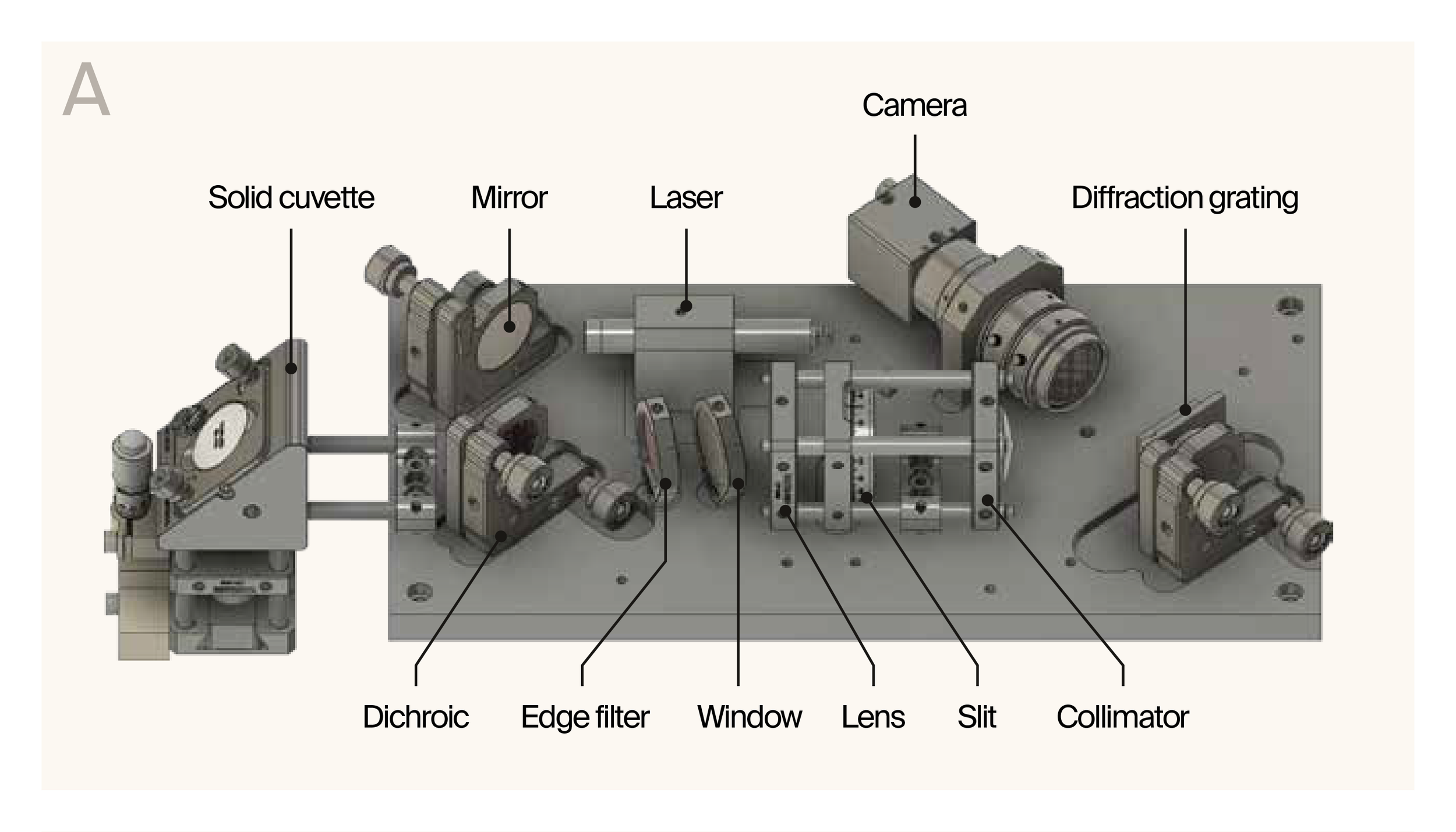
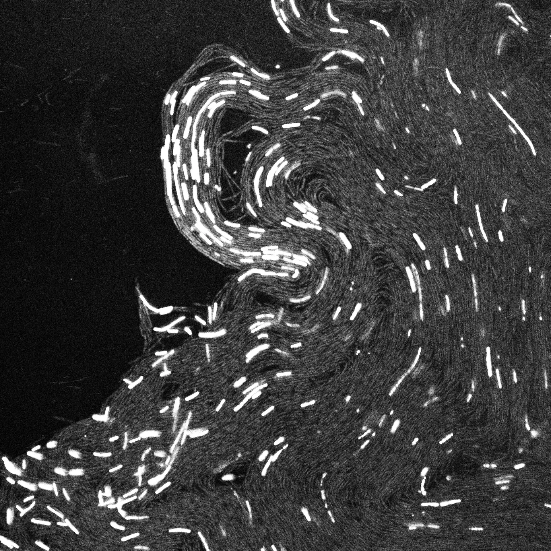
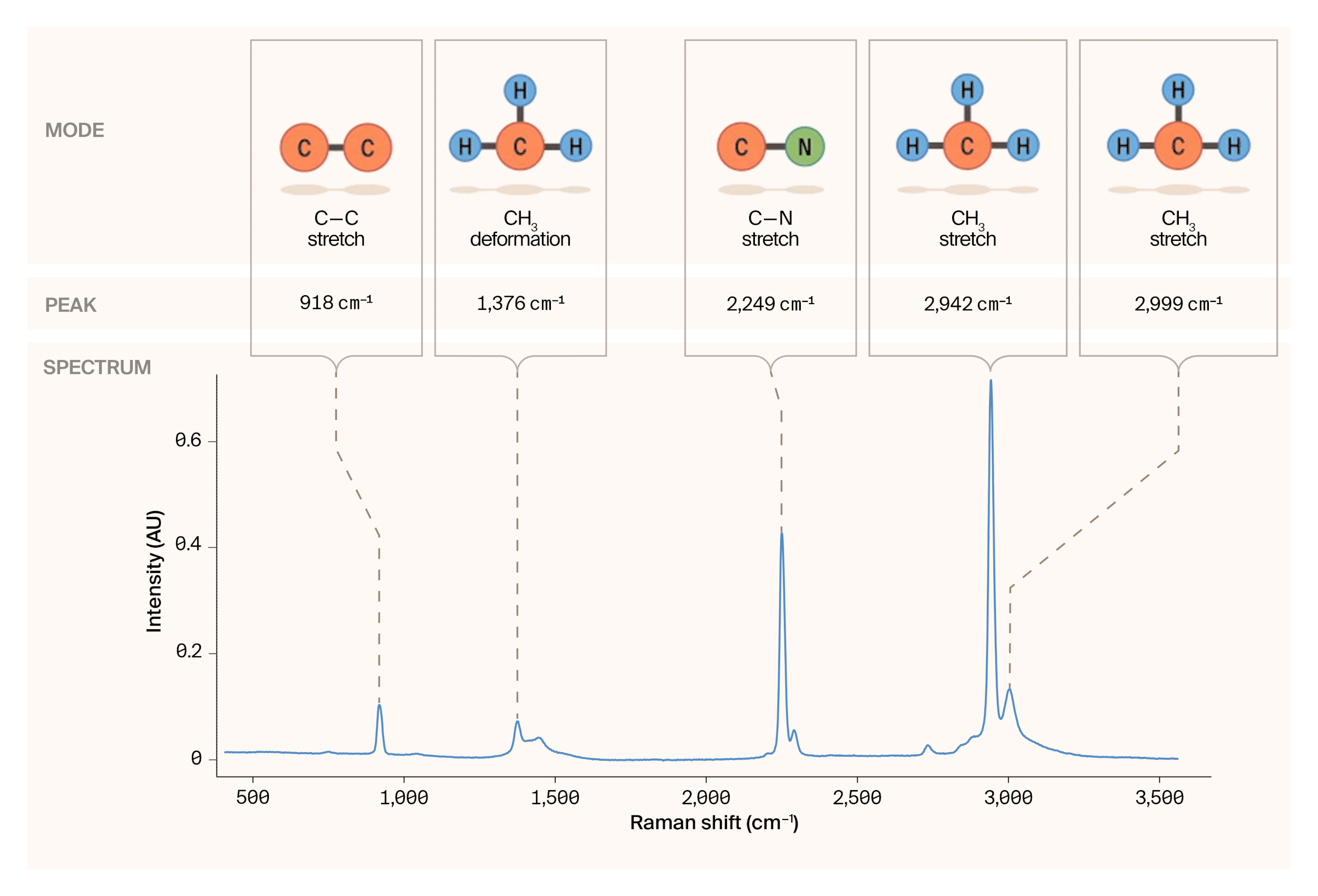
At Arcadia Science, I built the company's Raman spectroscopy capability from scratch. The starting point was a broken open-source instrument and no spectroscopy expertise in the company. I diagnosed and repaired the instrument, then designed a quantitative head-to-head evaluation of commercial Raman systems, leading to the procurement of two production systems. I now run high-throughput Raman phenotyping with bacteria, archaea, fungi, and protists, as well as protein structure characterization. I am also investigating the comparative utility of different spectroscopy and microscopy modalities and their analysis methods for explaining versus predicting biological phenomena. All work is published openly on thestacks.org.
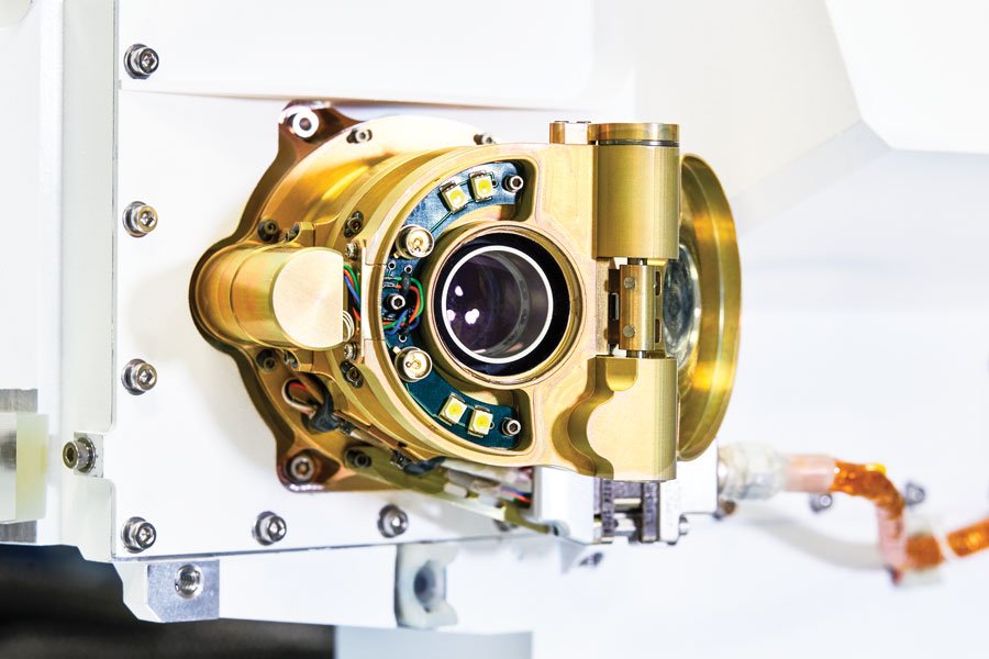
As the main operator of the two SHERLOC flight analog instruments at the Jet Propulsion Laboratory, I developed manuals for operation and troubleshooting, pipelines for data calibration and processing, and trained postdocs, students, and technologists. I established a standardization procedure, data structure, and organization for the analog data from our instruments and others on the team, which is now hosted on the Open Data Repository. I also contributed to publishing our analog spectral library, focusing on organic standards.
By using one of our flight analog instruments and developing a new set of experiments, I contributed to recovering the SHERLOC instrument back to operational status after it was taken offline. This feat (achieved from ~140 million miles away) was recognized with a NASA Award in 2024 for the SHERLOC Anomaly Recovery Team.
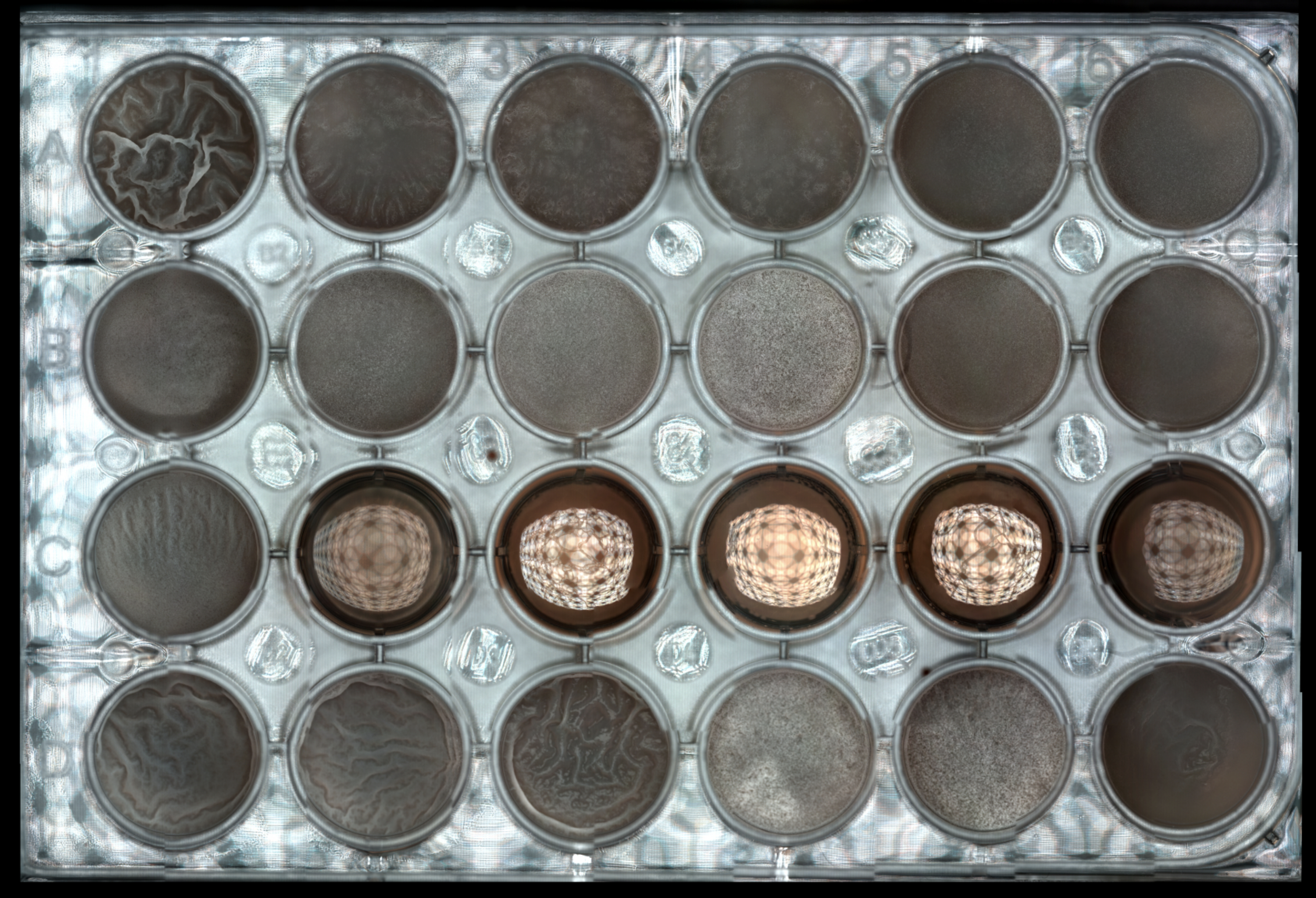
As a contracted research scientist through ex situ bio, I demonstrated new biological research applications for Ramona Optics' multi-camera array microscope (MCAM), a high-throughput gigapixel imaging system. This work resulted in three peer-reviewed publications, including a paper in Optica and one in eLife. I developed acquisition and analysis workflows for bacterial colony phenotyping at scale.
My introduction to scientific instrumentation was through microscopy and imaging. During graduate school at MIT, I designed microimaging, video, and timelapse setups to monitor cell growth, biochemical reactions, and animal movement, and applied computer vision for tracking and quantification. At the Institut de la Vision in Paris, I developed algorithms for detecting sister cells in confocal image stacks. At the Boyden Lab at MIT, I built optical and electrophysiological recording systems for awake behaving mice, including VR-based behavioral assays. I've worked with widefield, fluorescence, confocal, and scanning electron microscopy, and have maintained and repaired optical microscopes throughout my career.